- Suture Practice
- Medical simulator manikin
- Skeleton model
- Torso model
- Anatomical model
- Dental Model
- Other
Product Catalog
Contact Us

or
+ 86-28-81024434
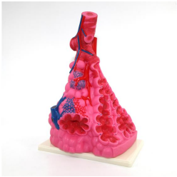
Alveolar Enlargement Model
SC3306-3
Description
1. This model is taken from the branch below the terminal trachea-the respiratory part. The model is about 400mm high and has a cone shape. It is installed on the bottom plate.
2. It shows the branch respiratory bronchioles, alveolar ducts, lung sacs and alveoli of terminal bronchiole.
3. A longitudinal section of one side of the alveolar duct shows alveolar hair (section) alveolar nodules and blood capillaries in the alveolar septum.
4. Make a cross-section of the alveolar sac on the other side, showing its cross-sectional structure.
5. Shows the smooth muscle, elastic fibers and reticular fibers surrounding the tube wall.
6. It shows the gradual branches of the pulmonary artery and pulmonary vein and the capillary network that surrounds the alveolar wall, and shows the bronchial arteries and veins.